This note discussed various neuroimaging methods used to image the Central Nervous System (CNS). These methods can be broadly categorized into two types:
- Structural Imaging: Creates an image of the anatomy of the brain, including any pathology or injury.
- Functional Imaging: Creates an image of the metabolic or pharmacologic activity of the brain, or cognitive functioning.
Neuroimaging techniques
-
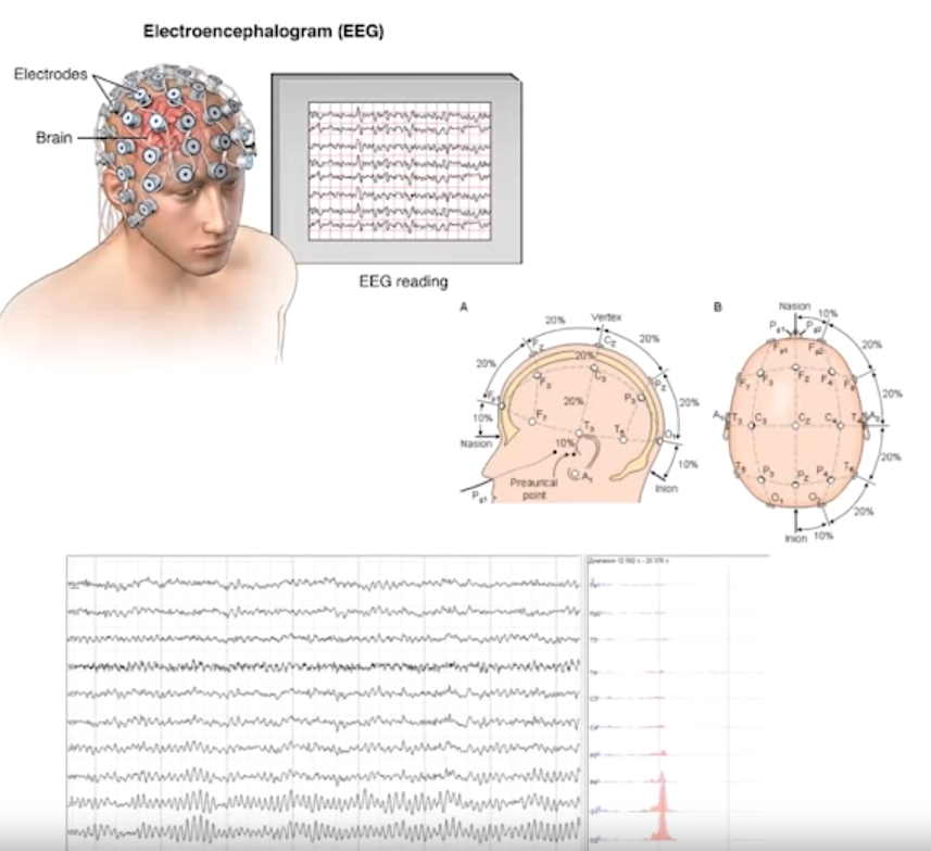
EEG (Electroencephalography): Measures electrical activity in the brain using electrodes placed on the scalp. Commonly used to study epilepsy and localize seizure onset.
-
-
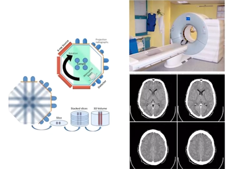
CT (Computed Tomography): Uses X-rays to create a 3D volumetric image of the brain. It’s fast, widely available, but involves radiation exposure.
-
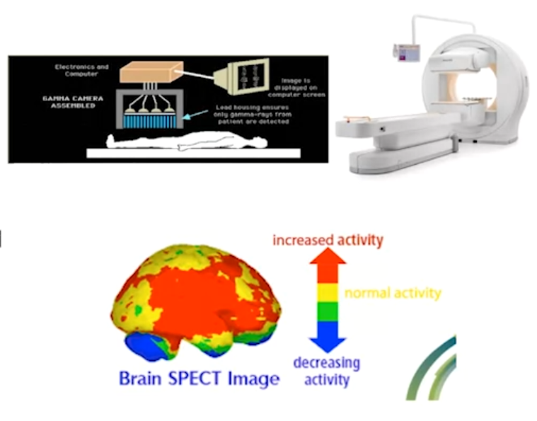
SPECT (Single-photon Emission Computed Tomography): A nuclear imaging technique that uses gamma-emitting tracers to measure brain activity. It has lower spatial resolution compared to other techniques.
-
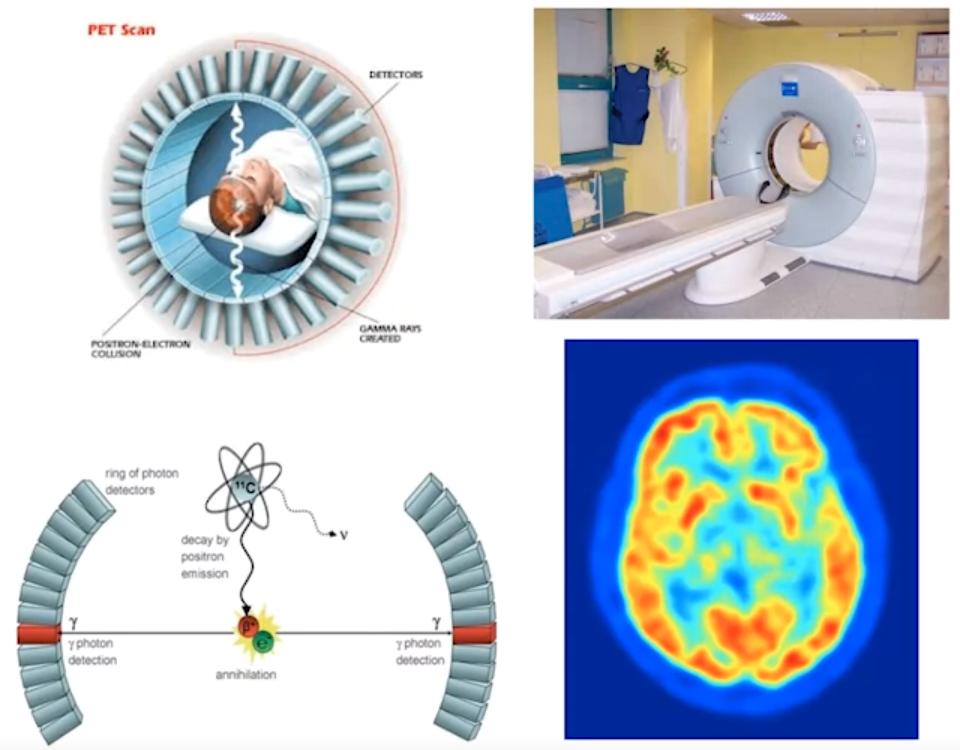
PET (Positron Emission Tomography): A refinement of SPECT, also uses gamma-emitting tracers but offers better spatial resolution. Commonly used for imaging glucose uptake (FDG-PET) and beta-amyloid plaques (18F-AV-45 PET) in Alzheimer’s disease.
-
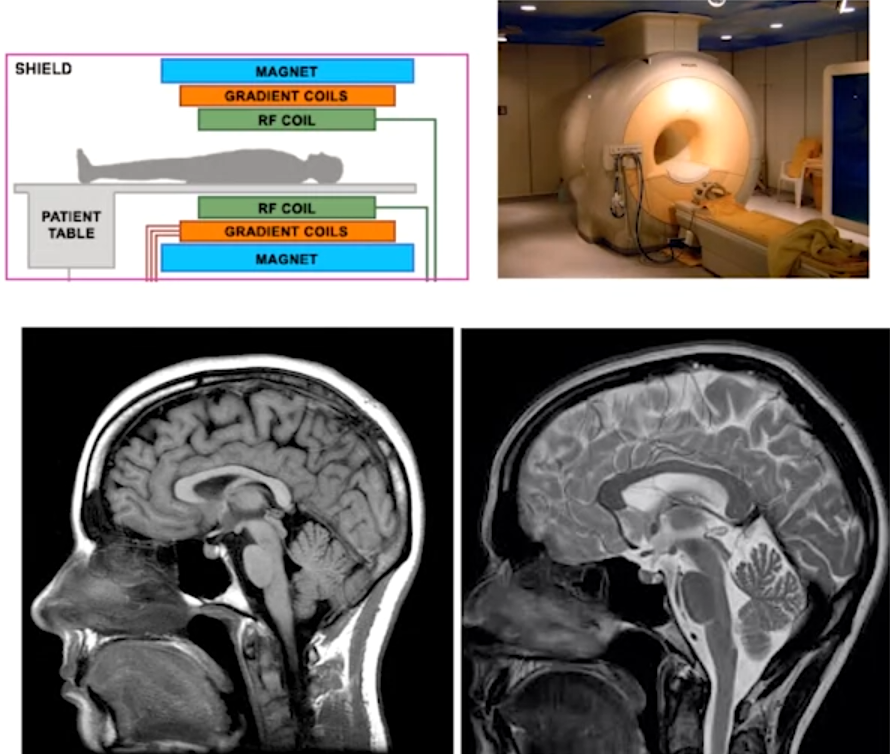
MRI (Magnetic Resonance Imaging): Uses strong magnetic fields and radio waves to create detailed images of the brain. It has good spatial resolution and safety for those who can undergo the procedure. MRI scanners use static magnetic fields, gradient coils, and radiofrequency coils to generate images. Functional MRI (fMRI) measures blood oxygenation levels to map brain activity during tasks. Diffusion Tensor Imaging (DTI) measures water diffusion to assess white matter integrity. Magnetic Resonance Spectroscopy (MRS) measures metabolite levels in specific brain regions.