I. fMRI Signal and BOLD Response
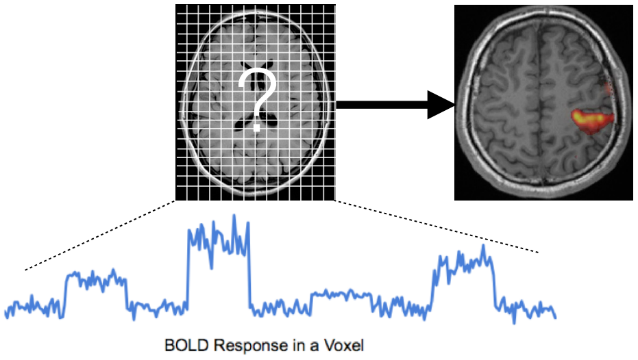
- Functional Magnetic Resonance Imaging (fMRI): Measures brain activity indirectly by detecting blood oxygen level changes.
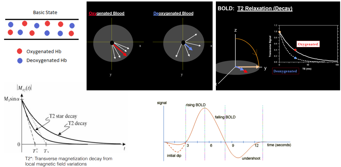
- Blood Oxygenation and Signal Strength: Oxygenated blood generates a stronger MRI signal compared to deoxygenated blood.
- T2 Relaxation Time:* Measures the time it takes for excited atoms to return to their resting state, sensitive to local magnetic field variations caused by blood oxygenation changes.
- T2 Decay and Neural Activity:* Lower T2* values due to increased oxygenation indirectly represent neural activity.
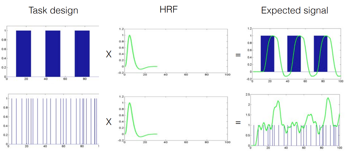
- Hemodynamic Response Function (HRF): Represents the relationship between neural activity and the corresponding blood flow changes.
II. Advantages of BOLD fMRI
- Non-invasive: Safe for human participants.
- High Spatial Resolution: Provides detailed information about brain activity location (around 1-1.5 millimeters).
- Good Temporal Resolution: Captures changes in brain activity relatively quickly (greater than half a second).
- Safety: Exposure to the magnetic field has no known long-lasting negative effects.
- Brain Network Analysis: Allows investigation of both localized activity in specific brain structures and interactions between different brain regions.
III. Designing an fMRI Experiment
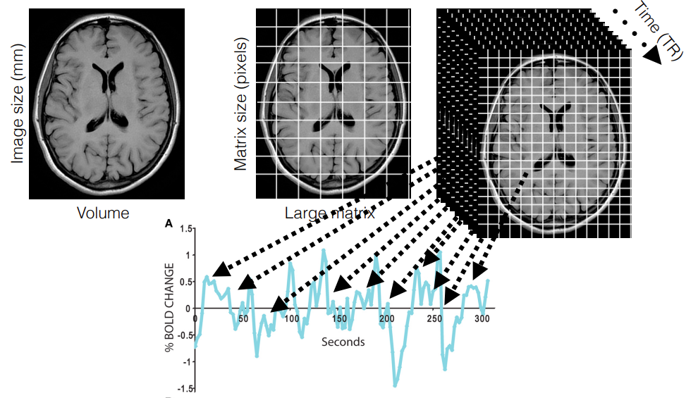
- Image Matrix Size: Determines the trade-off between spatial resolution (detail) and data acquisition time. A larger matrix captures more detail but takes longer to acquire.
- Time Series Data: T2* decay is measured repeatedly for each voxel (3D picture element) over time, resulting in a time series representing activation levels.
IV. Block Design
- Example: Finger Tapping Experiment
- Conditions: Rest (off) and finger tapping (on).
- Activation Time Series: Shows peaks corresponding to tapping periods, indicating higher activation during tapping.
- Motor Cortex: The cluster of activated voxels in the motor cortex is associated with finger tapping.
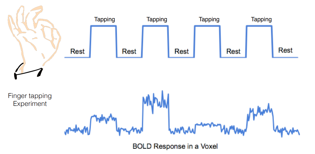
- Simple Design: Alternating blocks of “on” (task) and “off” (rest) conditions.
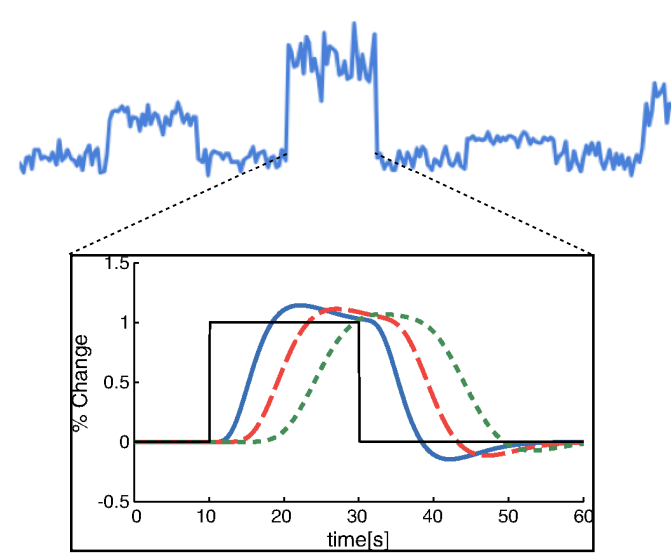
- Robust Activation: Well-suited for detecting sustained activation due to the accumulation of hemodynamic responses within a block.
- HRF Insensitivity: Less affected by variations in the shape or timing of the HRF.
- Limitations:
- Assumes Constant Activity: Requires a constant level of activation throughout the block.
- Limited Event Analysis: Cannot analyze individual events within a block, only overall block activation.
- HRF Variations Not Studied: Does not account for potential variations in the HRF across the brain.
 Up → block design | bottom → event design
V. Event-Related Design
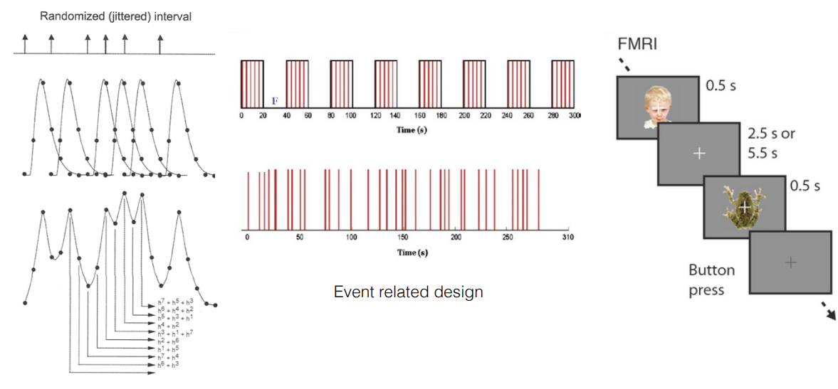
- Short Stimuli: Uses brief and separated stimulus presentations instead of continuous blocks.
- Individual Event Responses: Allows researchers to make inferences about the brain’s response to each individual event.
- HRF Sensitivity: More susceptible to variations in the HRF, which can affect the analysis.
- Advantages:
- Timing of Activation: Enables studying the timing of neural activation in response to specific events.
- Flexible Analysis: Allows for flexible sorting and analysis of trials after data collection.
- Disadvantages:
- Lower Power: Typically has lower statistical power to detect activation compared to block designs.
- HRF Dependence: Results can be significantly impacted by variations in the HRF.
- Carry-over Effects: Responses from one event might influence responses to subsequent events.
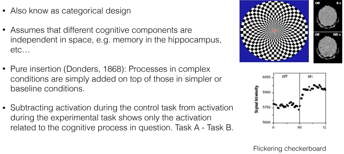
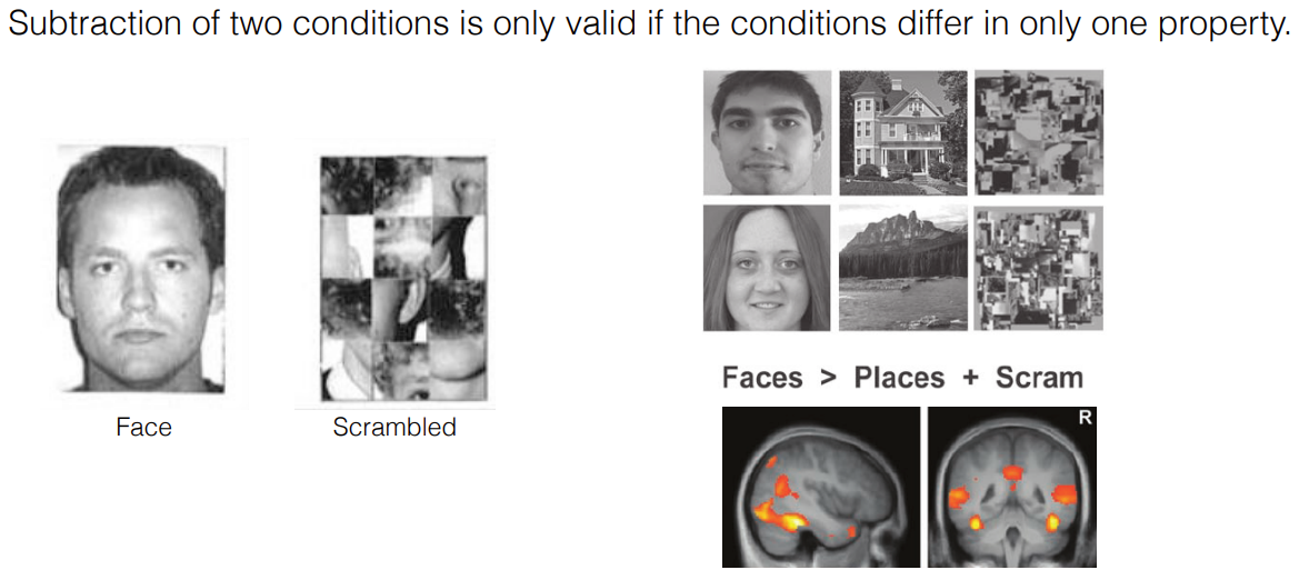
VI. Cognitive Subtraction Method
-
Contrasting Activation: Compares brain activity during a task condition with a control condition to isolate the brain activity related to the cognitive process of interest.
-
Assumptions: Relies on the assumptions that different cognitive processes are localized in distinct brain regions and that complex tasks can be understood as simpler tasks combined.
-
Control Condition Design: Careful design of the control condition is crucial to isolate the specific cognitive process under investigation. Resting baseline is generally a poor control due to ongoing mental activity (mind-wandering). A more effective approach is to use an active control condition that resembles the task condition but lacks the process of interest.
 VII. Example fMRI Experiments
-
Visual Perception: Investigates brain regions involved in vision by comparing brain activity during viewing a flickering checkerboard pattern to scrambled patterns.
-
Face Recognition: Identifies the brain region responsible for face processing by contrasting brain activity during viewing faces with scrambled faces and object pictures.
Key Takeaways:
- BOLD fMRI measures neural activity indirectly through blood oxygenation changes.
- The choice of experimental design (block vs. event-related) impacts the type of brain activity that can be effectively measured.
- Cognitive subtraction using a well-designed control condition is essential to isolate brain activity associated
[! Additional Resources] https://www.youtube.com/watch?v=avsYY5dbJ7g https://www.youtube.com/watch?v=RQrtNVKvIIk