I. Introduction
- The lecture dives into the sympathetic and parasympathetic nervous systems.
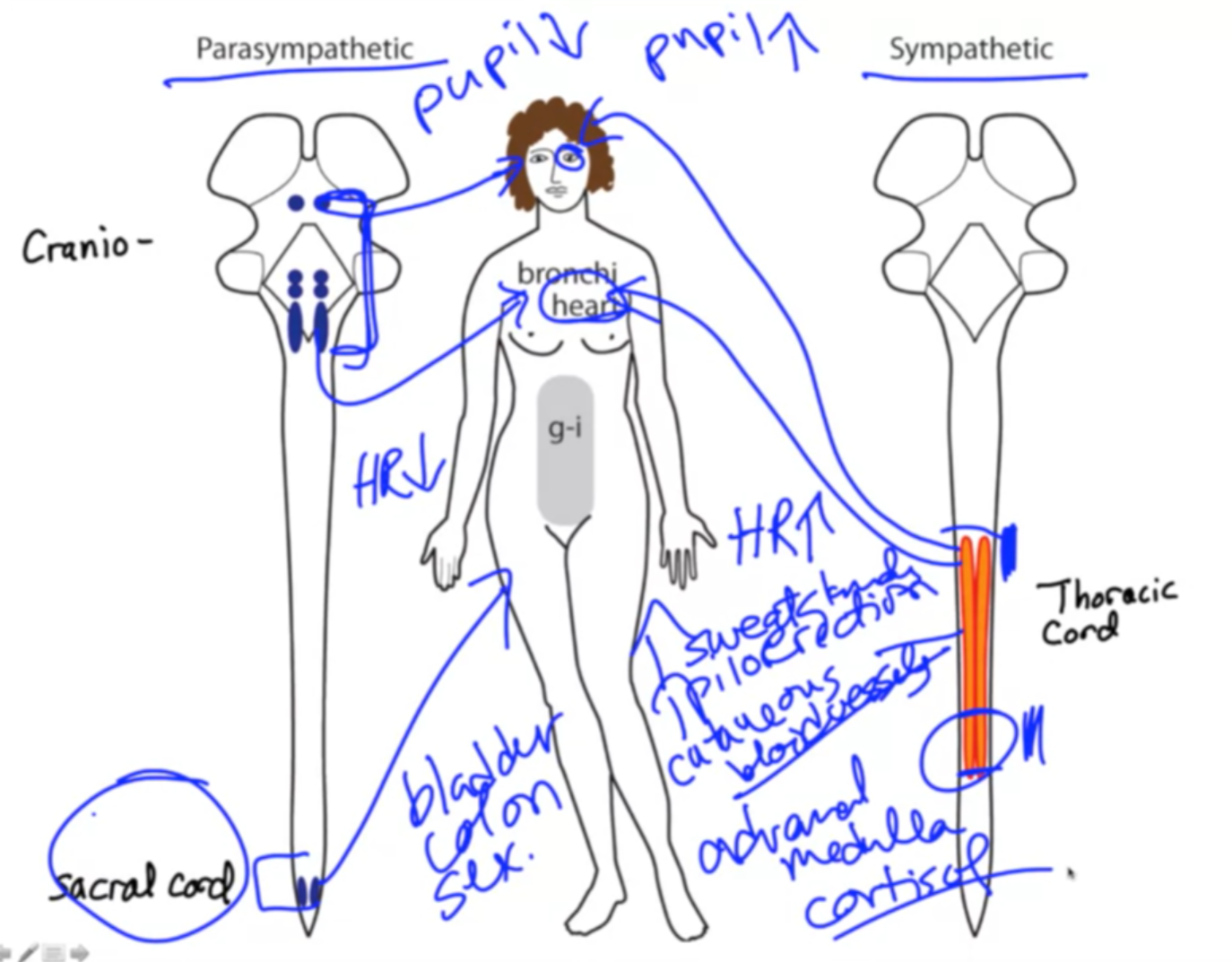
- Both are automatic and target three tissue types: smooth muscle, cardiac muscle, and glands.
II. Key Differences
- Function:
- Sympathetic: “Fight or flight” response (increased heart rate, pupil dilation, etc.)
- Parasympathetic: “Rest and digest” response (decreased heart rate, pupil constriction, etc.)
- Origin:
- Sympathetic: Thoracic spinal cord
- Parasympathetic: Cranial nerves and sacral spinal cord (often called craniosacral system)
III. Examples of Opposing Actions
- Heart: Sympathetic increases heart rate, parasympathetic decreases it.
- Eyes: Sympathetic dilates pupils, parasympathetic constricts them.
IV. Exceptions to the “Opposing Actions” Rule
- Sacral parasympathetics and sympathetics in the bladder, colon, and sexual organs:
- Bladder and colon: Both systems have roles, but parasympathetic is crucial for elimination.
- Sexual organs: Parasympathetic is important for arousal, while sympathetic is critical for climax.
V. Unique Functions of the Sympathetic System
- Sweat glands: Only innervated by the sympathetic system, responsible for sweating.
- Piloerection (goosebumps): Exclusively controlled by the sympathetic system.
- Cutaneous blood vessels: Sympathetic system controls constriction and dilation, impacting blood pressure.
- Adrenal medulla: Releases the stress hormone cortisol, solely innervated by the sympathetic system.
VI. Clinical Example: Addison’s Disease
- Caused by insufficient adrenal medulla function, leading to cortisol deficiency.
- Symptoms include fatigue, weakness, and weight loss.
- John F. Kennedy is a historical example of someone with Addison’s disease.
VII. Conclusion
- The sympathetic and parasympathetic systems work together to maintain physiological balance.
- Understanding their individual functions and interactions is crucial in various physiological contexts. Sympathetic Balance
Key points:
- The parasympathetic preganglionic neurons are located in the brainstem and sacral cord, while the sympathetic preganglionic neurons are located in the thoracic cord.
- Spinal cord injury can isolate the sacral cord, which is responsible for bladder function.
- When the sacral cord is isolated, it can no longer receive instructions from the brain and may cause bladder dyssynergia, a condition where the bladder contracts while the sphincter remains closed.
- Bladder dyssynergia is a common medical emergency following spinal cord injury.
Additional details:
- The sacral cord is responsible for executing bladder function, but it does not make the decision to void urine.
- Micturition (urination) requires both the contraction of the bladder (a parasympathetic effect) and the relaxation of the sphincter (a voluntary muscle controlled by the brain).
- In spinal cord injury, the bladder may contract but the sphincter will not relax due to the lack of communication between the brain and the sacral cord.
- This can lead to serious health problems if not addressed promptly.
Interesting fact:
- Eyelid movement can be used to assess wake-sleep cycles in individuals with spinal cord injury because the eyelid is controlled by the sympathetic nervous system, which remains intact in some cases.